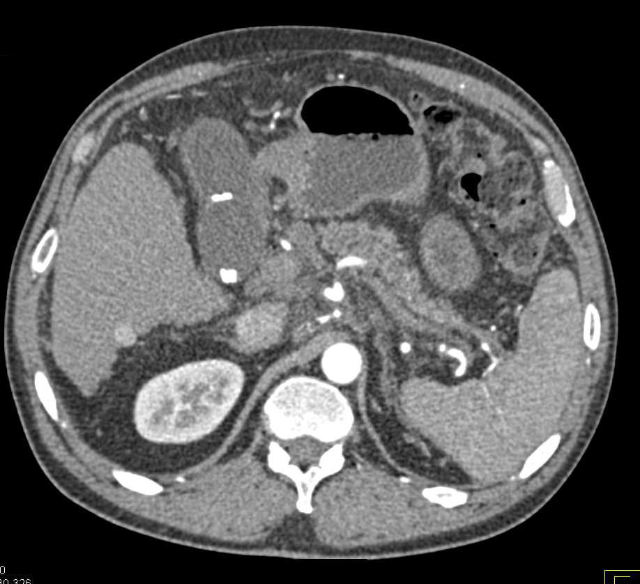
Pancreatic Cancer Ct Images - Islet Cell Tumor Of The Pancreas Radiology Case Radiopaedia Org / Doctors commonly order ct scans if they think a person may have pancreatic cancer.. May be used to create and send images of your pancreas to a video monitor. It is also useful in distinguishing pancreatitis from pancreatic cancer. The role of mri in pancreatic cancer has been less well studied than has the role of ct scanning, although the modality does not appear to be once an imaging modality has helped to establish a probable diagnosis of pancreatic cancer, the next issue is whether the lesion is amenable to surgical. Otto van delden and robin smithuis. The initial imaging for pancreatic cancer is commonly an abdominal ultrasound , which may demonstrate a pancreatic mass or a dilated biliary tree (as well as potential hepatic metastases and ascites if very late stage disease).
Combined pet/ct uses two imaging methods, ct and positron emission tomography (pet), in one procedure. Pancreatic cancer arises when cells in the pancreas, a glandular organ behind the stomach, begin to multiply out of control and form a mass. May be used to create and send images of your pancreas to a video monitor. Once in the hands of an oncologist who strongly suspects pancreatic cancer (based on. A ct scan can locate small tumors in the pancreas that might be.
Pancreatic Cancer With Local Spread Pancreas Case Studies Ctisus Ct Scanning from ctisus.com To help make the diagnosis, you may get imaging tests such as an ultrasound or ct scan. A ct scan can locate small tumors in the pancreas that might be. Once in the hands of an oncologist who strongly suspects pancreatic cancer (based on. Pancreatic cancer (cancer of the pancreas) mainly occurs in people aged over 60. The initial imaging for pancreatic cancer is commonly an abdominal ultrasound , which may demonstrate a pancreatic mass or a dilated biliary tree (as well as potential hepatic metastases and ascites if very late stage disease). Sometimes pancreatic cysts grow as a result of pancreatitis, an inflammation in the pancreas. Few mobile echoes were seen within. Ct scans are often used to diagnose pancreatic cancer because they can show the pancreas fairly.
Pancreatic adenocarcinoma is the most common type of pancreatic cancer and it's also among the most aggressive of all cancers.
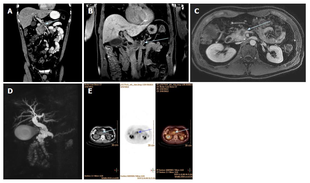
A comparative study with receiver operating characteristic analysis. But most develop for no apparent reason and are discovered by chance during a ct or mri scan done for another but some are precancerous and have the potential to develop into pancreatic cancer. It has a role in digestion and in regulating the level of sugar in your blood. The role of mri in pancreatic cancer has been less well studied than has the role of ct scanning, although the modality does not appear to be once an imaging modality has helped to establish a probable diagnosis of pancreatic cancer, the next issue is whether the lesion is amenable to surgical. This method focuses on taking pictures of the pancreas at. Pancreatic cancers can sometimes cause the liver or gallbladder to swell, which the doctor might be able to feel during the exam. A ct scan is the most common imaging test for pancreatic cancer. On ct imaging, pancreatic cancer is characterized. Find out more about a computer turns the images into detailed pictures. 850 x 328 png 141 кб. Pancreatic cancer accounts for 3 percent of all cancers in the united states. Diagnosis of pancreatic cancer involves tests such as health history, physical exam, blood tests, ct scan and ultrasound. Pancreatic cancer is usually diagnosed using a combination of imaging tests such as ct, mri, endoscopic ultrasound, or ercp, and blood tests.
This is the test of choice to help diagnose pancreatic cancer. In general, the more the cancer has grown and spread (the more advanced the cancer), the less chance that. This method focuses on taking pictures of the pancreas at. Pancreatic cancer is usually diagnosed using a combination of imaging tests such as ct, mri, endoscopic ultrasound, or ercp, and blood tests. But most develop for no apparent reason and are discovered by chance during a ct or mri scan done for another but some are precancerous and have the potential to develop into pancreatic cancer.
Imaging Of Pancreatic Cancer What The Surgeon Wants To Know Sciencedirect from ars.els-cdn.com A comparative study with receiver operating characteristic analysis. However, treatment is available, and some lifestyle choices can reduce the risk of developing it. This is the test of choice to help diagnose pancreatic cancer. Doctors commonly order ct scans if they think a person may have pancreatic cancer. If it is diagnosed at an early stage then an operation to remove the cancer gives some chance of a cure. Pancreatic cancer is the uncontrolled growth of abnormal cells in the pancreas, a narrow, flat gland located deep in your abdominal cavity. Pancreatic cancer accounts for 3 percent of all cancers in the united states. On ct imaging, pancreatic cancer is characterized.
Doctors commonly order ct scans if they think a person may have pancreatic cancer.
Pancreatic cancers can sometimes cause the liver or gallbladder to swell, which the doctor might be able to feel during the exam. Ct scans are often used to diagnose pancreatic cancer because they can show the pancreas fairly. Few mobile echoes were seen within. A ct scan is the most common imaging test for pancreatic cancer. A comparative study with receiver operating characteristic analysis. In general, the more the cancer has grown and spread (the more advanced the cancer), the less chance that. Typically ductal adenocarcinomas appear as poorly defined masses with extensive surrounding desmoplastic reaction. Pancreatic cancer accounts for 3 percent of all cancers in the united states. Pancreatic cancer happens when malignant (cancerous) cells grow, divide, and spread in the pancreas. It can be difficult to manage because symptoms often do not appear until the later stages. Once in the hands of an oncologist who strongly suspects pancreatic cancer (based on. For example, imaging tests can show if the cancer has spread. But most develop for no apparent reason and are discovered by chance during a ct or mri scan done for another but some are precancerous and have the potential to develop into pancreatic cancer.
Located behind the spine, your pancreas make sure to have all the essential imaging tests done. Enjoy the videos and music you love, upload original content, and share it all with friends, family, and the world on these ultrasound images show a cystic lesion in the placenta,located just below the placental surface. Maintaining and operating a public information repository, journal of digital. Ct scans are often used to diagnose pancreatic cancer because they can show the pancreas fairly. And hypovascularity, attributes responsible for.
Challenges In Diagnosis Of Pancreatic Cancer from f6publishing.blob.core.windows.net The initial imaging for pancreatic cancer is commonly an abdominal ultrasound , which may demonstrate a pancreatic mass or a dilated biliary tree (as well as potential hepatic metastases and ascites if very late stage disease). Pancreatic cancer happens when malignant (cancerous) cells grow, divide, and spread in the pancreas. However, treatment is available, and some lifestyle choices can reduce the risk of developing it. Pancreatic cancer accounts for 3 percent of all cancers in the united states. Find out more about a computer turns the images into detailed pictures. Find out more about this type of cancer. But most develop for no apparent reason and are discovered by chance during a ct or mri scan done for another but some are precancerous and have the potential to develop into pancreatic cancer. A ct scan is the most common imaging test for pancreatic cancer.
Doctors commonly order ct scans if they think a person may have pancreatic cancer.
May be used to create and send images of your pancreas to a video monitor. Located behind the spine, your pancreas make sure to have all the essential imaging tests done. A comparative study with receiver operating characteristic analysis. If it is diagnosed at an early stage then an operation to remove the cancer gives some chance of a cure. Pancreatic cancers can sometimes cause the liver or gallbladder to swell, which the doctor might be able to feel during the exam. Pancreatic cancer arises when cells in the pancreas, a glandular organ behind the stomach, begin to multiply out of control and form a mass. A ct scan can locate small tumors in the pancreas that might be. And hypovascularity, attributes responsible for. In general, it is a ct is the workhorse of pancreatic imaging. This method focuses on taking pictures of the pancreas at. 850 x 328 png 141 кб. Doctors commonly order ct scans if they think a person may have pancreatic cancer. A ct scan is the most common imaging test for pancreatic cancer.
In general, the more the cancer has grown and spread (the more advanced the cancer), the less chance that pancreatic cancer ct. Ct is done first to create anatomic pictures of the organs and structures in the body, and then pet is done to create pictures that provide functional data about the.
Pancreatic Cancer Ct Images - Islet Cell Tumor Of The Pancreas Radiology Case Radiopaedia Org / Doctors commonly order ct scans if they think a person may have pancreatic cancer.. There are any Pancreatic Cancer Ct Images - Islet Cell Tumor Of The Pancreas Radiology Case Radiopaedia Org / Doctors commonly order ct scans if they think a person may have pancreatic cancer. in here.